
Cases From the Clinic: A Non-Healing Wound of the Throatlatch of a Mare
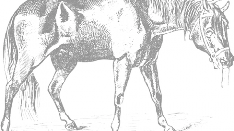
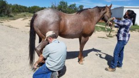
Bella came to our veterinary practice with a long-standing,
non-healing wound of her right throatlatch.
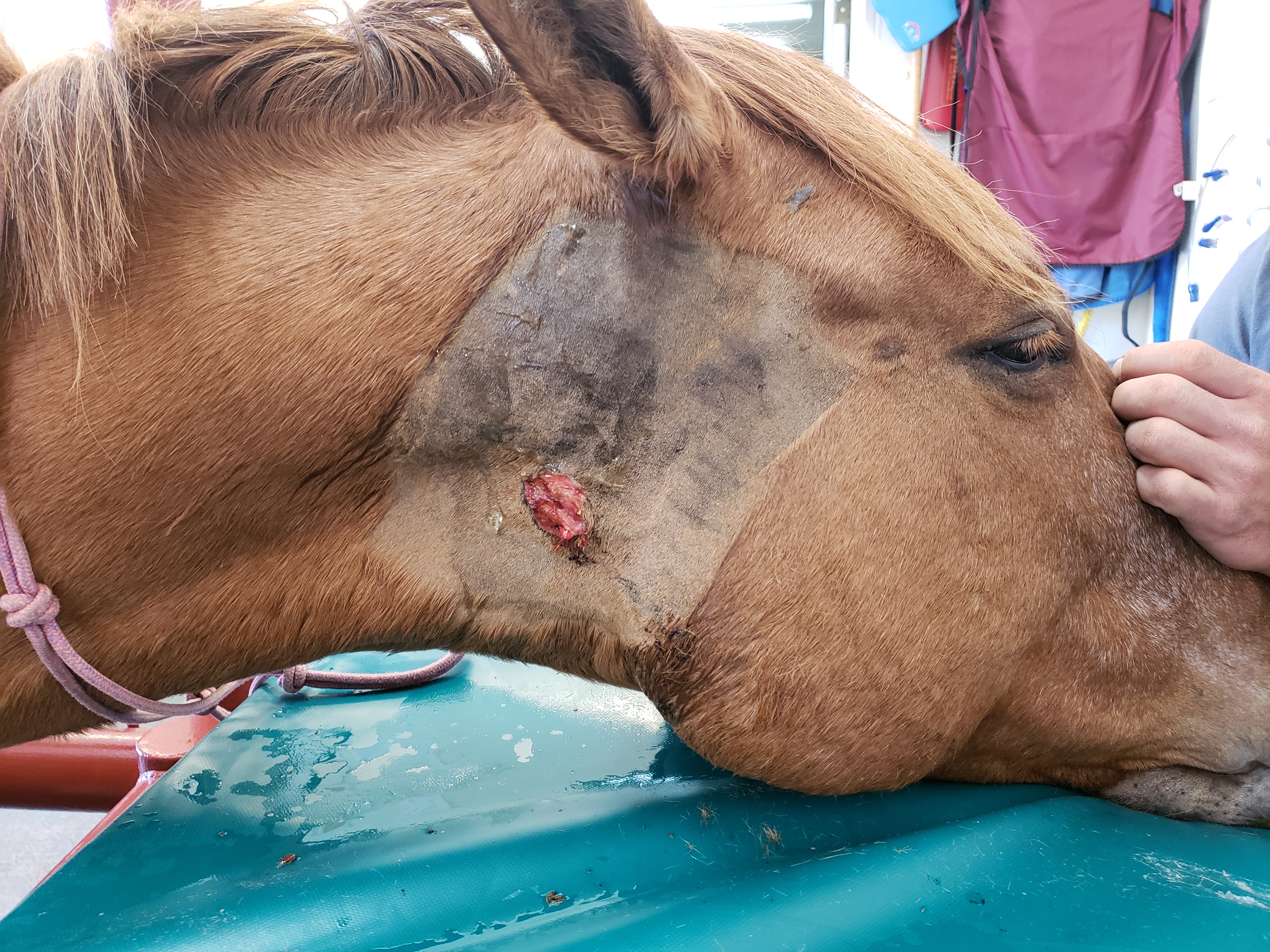
Bella, sedated and with the wound cleaned up.
History: The wound had been there over 6 months, and to the owner’s knowledge she didn’t have an accident that caused it. Over that time, she had been to a few different veterinarians and had been on antibiotic courses, wound cleaning and flushing. But the wound persisted.
Examination: She seemed well otherwise, her physical exam was normal. She had not lost weight. She had no cough or nasal discharge.
So let me ask you, how would you expect your vet to proceed with this?
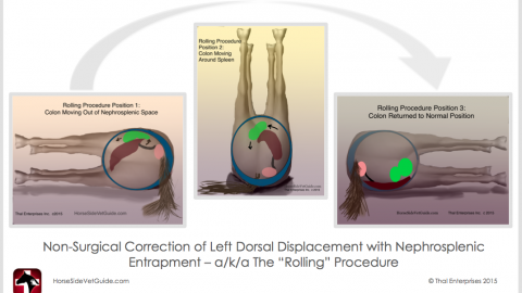
First off, it is really important to know the general anatomy of an area. Here are (some) of the important structures under the skin of the neck. A vet wants to be sure these structures aren’t affected. You would have to be really careful opening up this area:
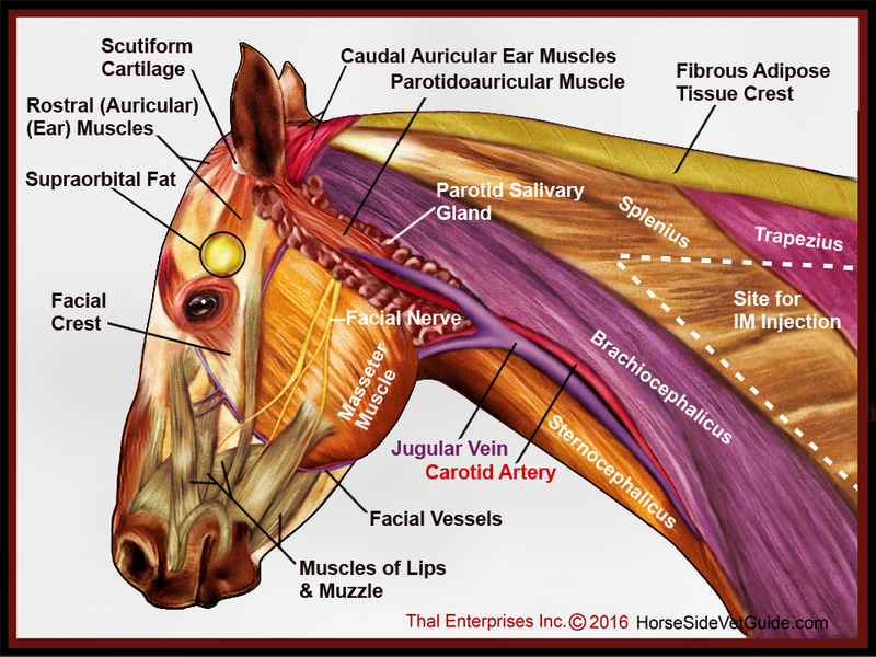
An image I created for Horse Side Vet Guide, that shows the main structures under the skin of the neck & head.
If you used Horse Side Vet Guide, you could educate yourself about that. I found this record “Wound is very slow healing or not healing.”
I gently cleaned and probed the wound to see the direction it went. It was obviously at least 4 inches deep, going up toward the base of the ear. I talked to the owner about the tests we could perform to try to diagnose the cause. I told him that radiographs would be a good place to start.
This radiograph (X-ray) shows a very interesting finding!
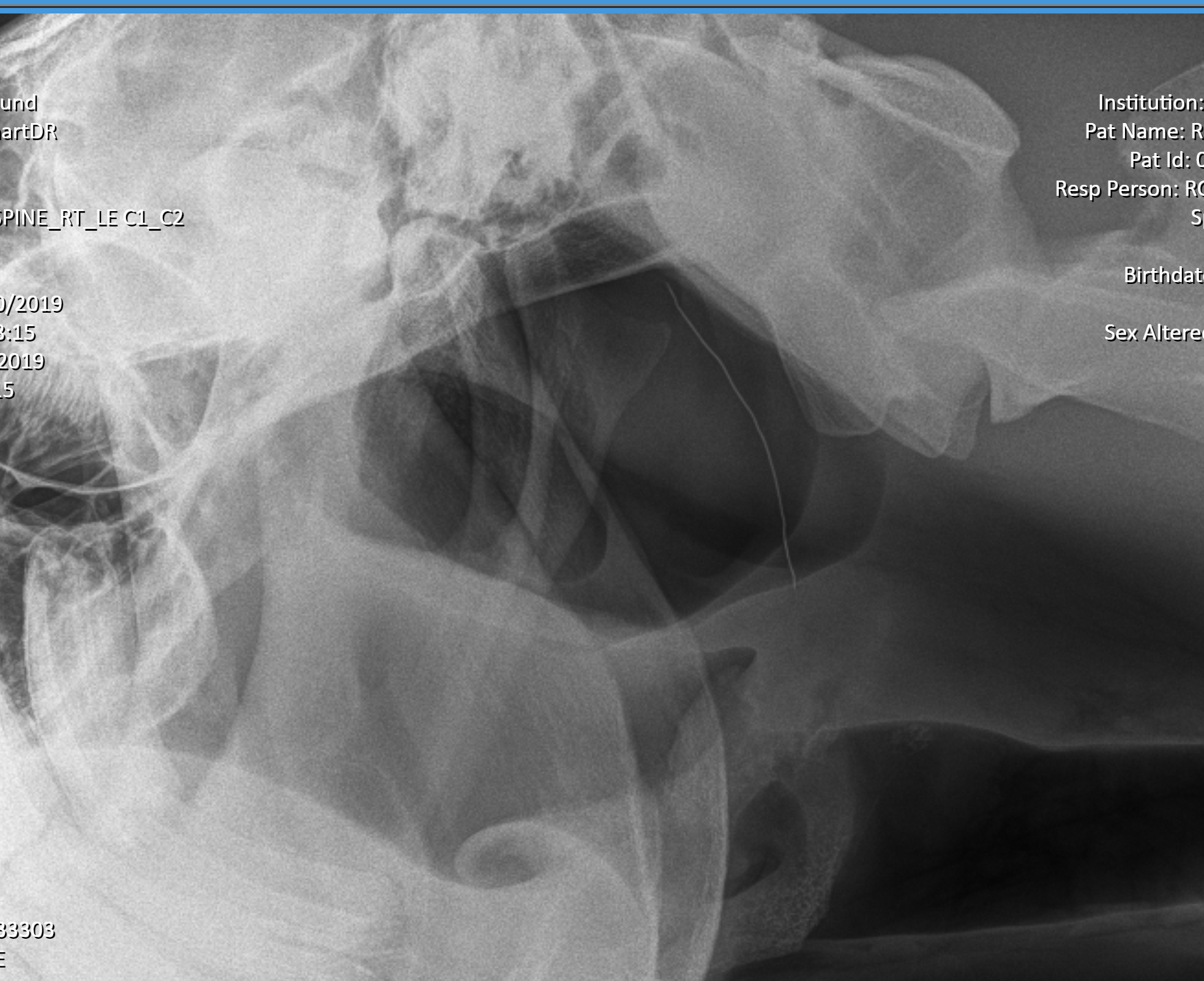
Look closely at this radiograph. The answer lies here!
As you can see, there is a thin (white) wire overlying the dark area in the center of the image (the guttural pouches). A foreign body was the reason the wound would not heal. The question is how it got there!
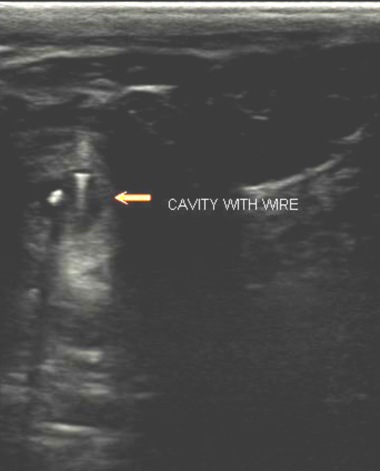
In these ultrasound images, the bright white speck is the wire, within a cavity that contains some air (second white spot). Ultrasound cannot penetrate metal or air. In this image, the metal is even brighter. The black “moat” around the wire is the draining tract cavity,
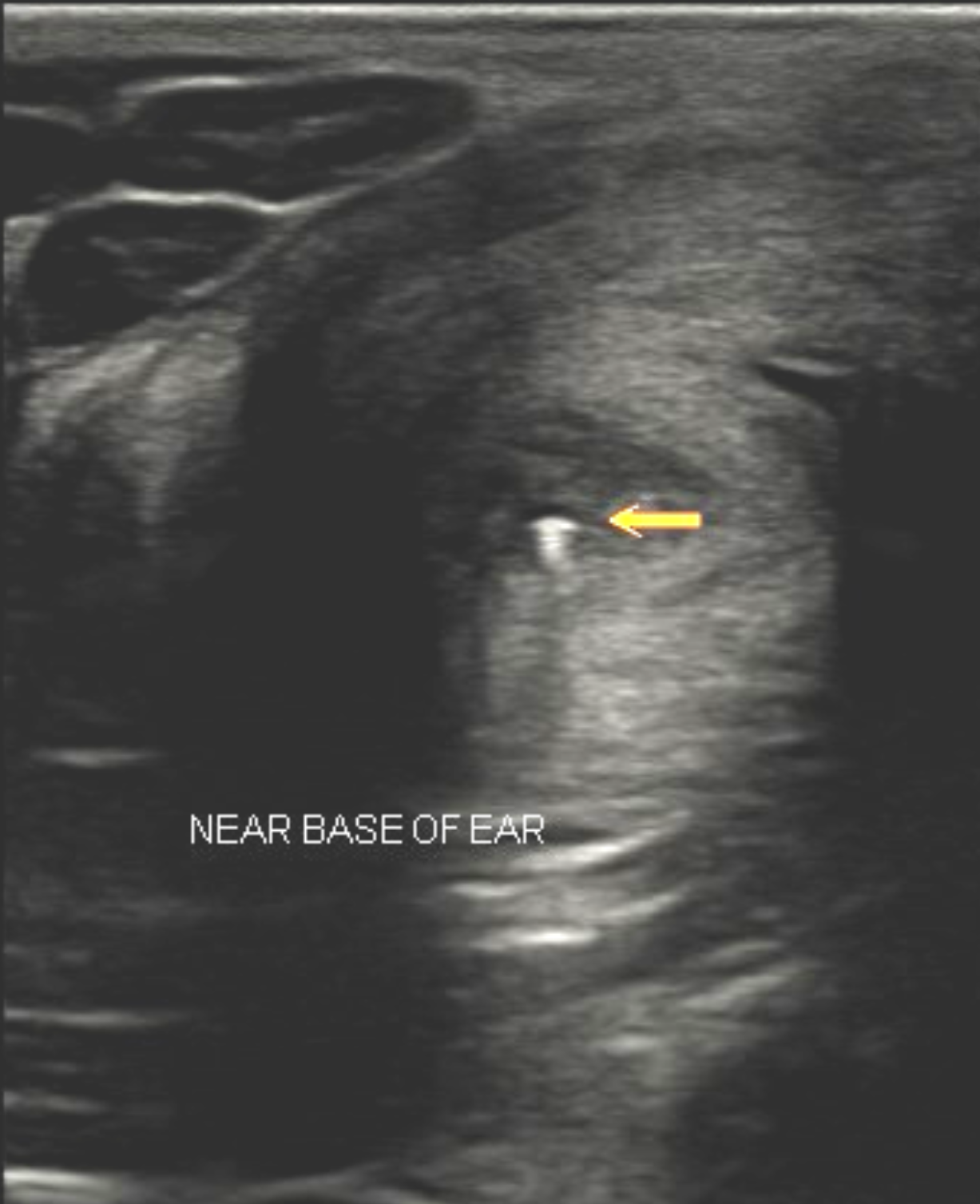
Ultrasound added to our understanding of the position of the wire. Here ultrasound showed us that the wire ran up along the side of the neck, about 3cm (1.5″) under the skin and toward the base of the ear. Under ultrasound guidance, I was able to pass a clamp up the tract and in real-time, grasp the wire and pull it out.
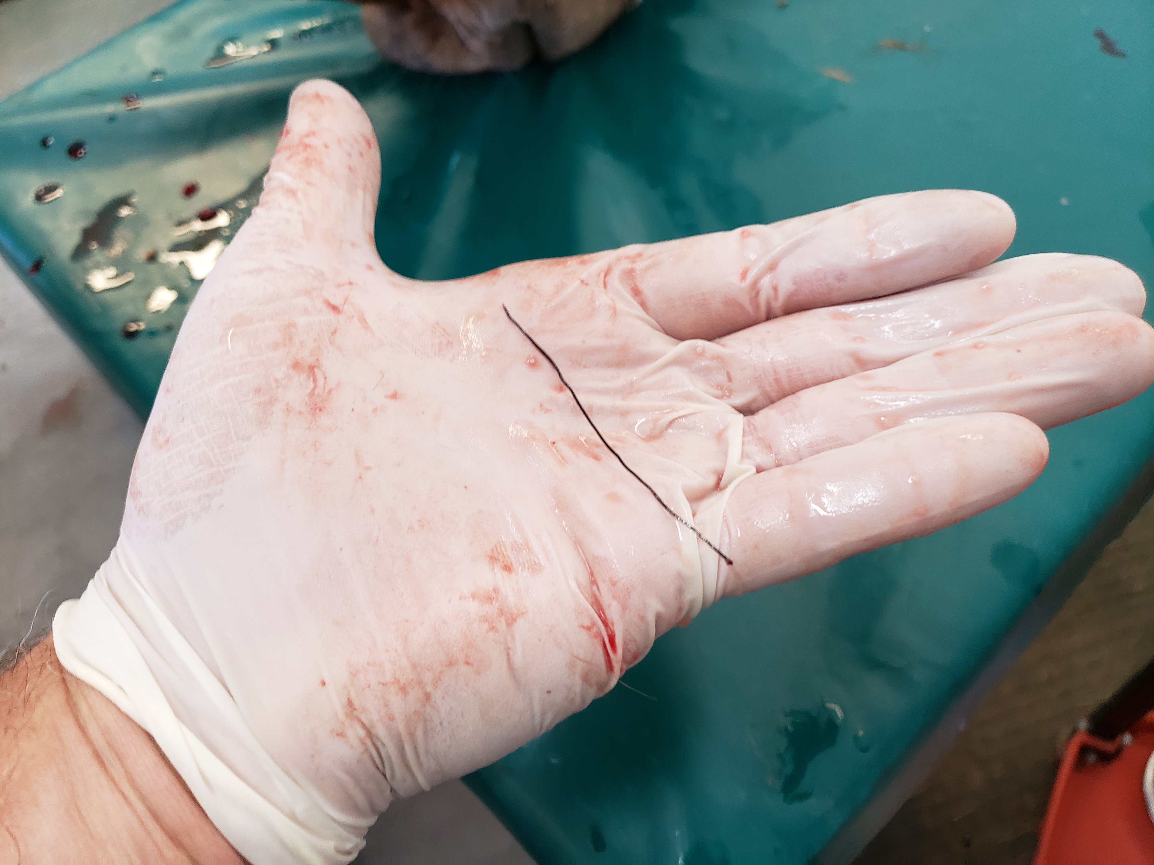
Treatment: Using the ultrasound, we were able to grasp the wire with a forceps and pull it out. I could not have done it without the ultrasound. It would have required that the whole area be opened up surgically and explored.
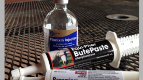
We started Bella on a course of antibiotic and so far things are healing well. Our hope is that simply getting that wire out of the tissues is enough to allow the wound to heal. I just spoke to the owner and suddenly the wound is healing a lot faster! One question that remains is where the wire came from. My best guess is that it was in her feed, it embedded itself in the tissues of the mouth, then migrated through the tissues to its present position.
-
Doug Thal DVM DABVP – Creator of Horse Side Vet Guide


